Hyaline Cartilage Drawing
Hyaline Cartilage Drawing - Step by step drawing of histology of. Replaced by bone tissue as the organ grows in length epiphysis wide section at each end. Web hyaline cartilage drawing. Tracheal cartilage, supporting connective tissue, 250x at 35mm. Web (also, growth plate) sheet of hyaline cartilage in the metaphysis of an immature bone; Web hyaline cartilage, the most abundant type of cartilage, plays a supportive role and assists in movement. Web how to draw histology of hyaline cartilage ? Formed by the process of chondrogenesis, the resulting. Web during embryonic development, hyaline cartilage serves as temporary cartilage models that are essential precursors to the formation of most of the axial and. Web hyaline cartilage a higher magnification of the wall of the trachea shows the lumen with its epithelial lining in the lower left of the image. Step by step drawing of histology of. Tracheal cartilage, supporting connective tissue, 250x at 35mm. A type of cartilage found on many joint surfaces; Web hyaline cartilage drawing. Most of the image is occupied by a section. Web how to draw histology of hyaline cartilage ? Web likecomment share subscribe #hyalinecartilage #histodiagrams #hyalinecartilagediagram #cartilagehistology Tracheal cartilage, supporting connective tissue, 250x at 35mm. Although hyaline cartilage feels nearly as hard and dense as bone. It contains no nerves or blood vessels, and its structure is relatively simple. Web how to draw histology of hyaline cartilage ? Web hyaline cartilage, the most abundant type of cartilage, plays a supportive role and assists in movement. Although hyaline cartilage feels nearly as hard and dense as bone. Web hyaline cartilage is a supportive connective tissue with a rigid yet slightly flexible extracellular matrix. Web hyaline cartilage drawing. A type of cartilage found on many joint surfaces; Web (also, growth plate) sheet of hyaline cartilage in the metaphysis of an immature bone; 5.4k views 3 years ago. Territorial matrix lies immediately around each. Most of the image is occupied by a section. Although hyaline cartilage feels nearly as hard and dense as bone. Web hyaline cartilage a higher magnification of the wall of the trachea shows the lumen with its epithelial lining in the lower left of the image. Web hyaline cartilage, the most abundant type of cartilage, plays a supportive role and assists in movement. Formed by the process of chondrogenesis,. Web hyaline cartilage a higher magnification of the wall of the trachea shows the lumen with its epithelial lining in the lower left of the image. Territorial matrix lies immediately around each. Web during embryonic development, hyaline cartilage serves as temporary cartilage models that are essential precursors to the formation of most of the axial and. It is also most. Web how to draw histology of hyaline cartilage ? A type of cartilage found on many joint surfaces; Step by step drawing of histology of. 5.4k views 3 years ago. Web hyaline cartilage is a supportive connective tissue with a rigid yet slightly flexible extracellular matrix. A type of cartilage found on many joint surfaces; Cartilage is one of the. It contains no nerves or blood vessels, and its structure is relatively simple. Step by step drawing of histology of. Web hyaline cartilage drawing. Replaced by bone tissue as the organ grows in length epiphysis wide section at each end. Step by step drawing of histology of. It is also most commonly found in the ribs, nose, larynx, and trachea. Cartilage is one of the. Web during embryonic development, hyaline cartilage serves as temporary cartilage models that are essential precursors to the formation of. Territorial matrix lies immediately around each. Isogenous groups and interstitial growth results when chondrocytes divide and produce extracellular matrix. Formed by the process of chondrogenesis, the resulting. Web this is joao from kenhub, and in this tutorial, we will be discussing the most common type of cartilage found in the human body which is hyaline cartilage. Web during embryonic development,. Most of the image is occupied by a section. Web this is joao from kenhub, and in this tutorial, we will be discussing the most common type of cartilage found in the human body which is hyaline cartilage. It contains no nerves or blood vessels, and its structure is relatively simple. Cartilage is one of the. Web likecomment share subscribe #hyalinecartilage #histodiagrams #hyalinecartilagediagram #cartilagehistology Isogenous groups and interstitial growth results when chondrocytes divide and produce extracellular matrix. Web (also, growth plate) sheet of hyaline cartilage in the metaphysis of an immature bone; Although hyaline cartilage feels nearly as hard and dense as bone. Web hyaline cartilage a higher magnification of the wall of the trachea shows the lumen with its epithelial lining in the lower left of the image. Web how to draw histology of hyaline cartilage ? 5.4k views 3 years ago. It is also most commonly found in the ribs, nose, larynx, and trachea. Tracheal cartilage, supporting connective tissue, 250x at 35mm. Web hyaline cartilage is a supportive connective tissue with a rigid yet slightly flexible extracellular matrix. Web hyaline cartilage drawing. Step by step drawing of histology of.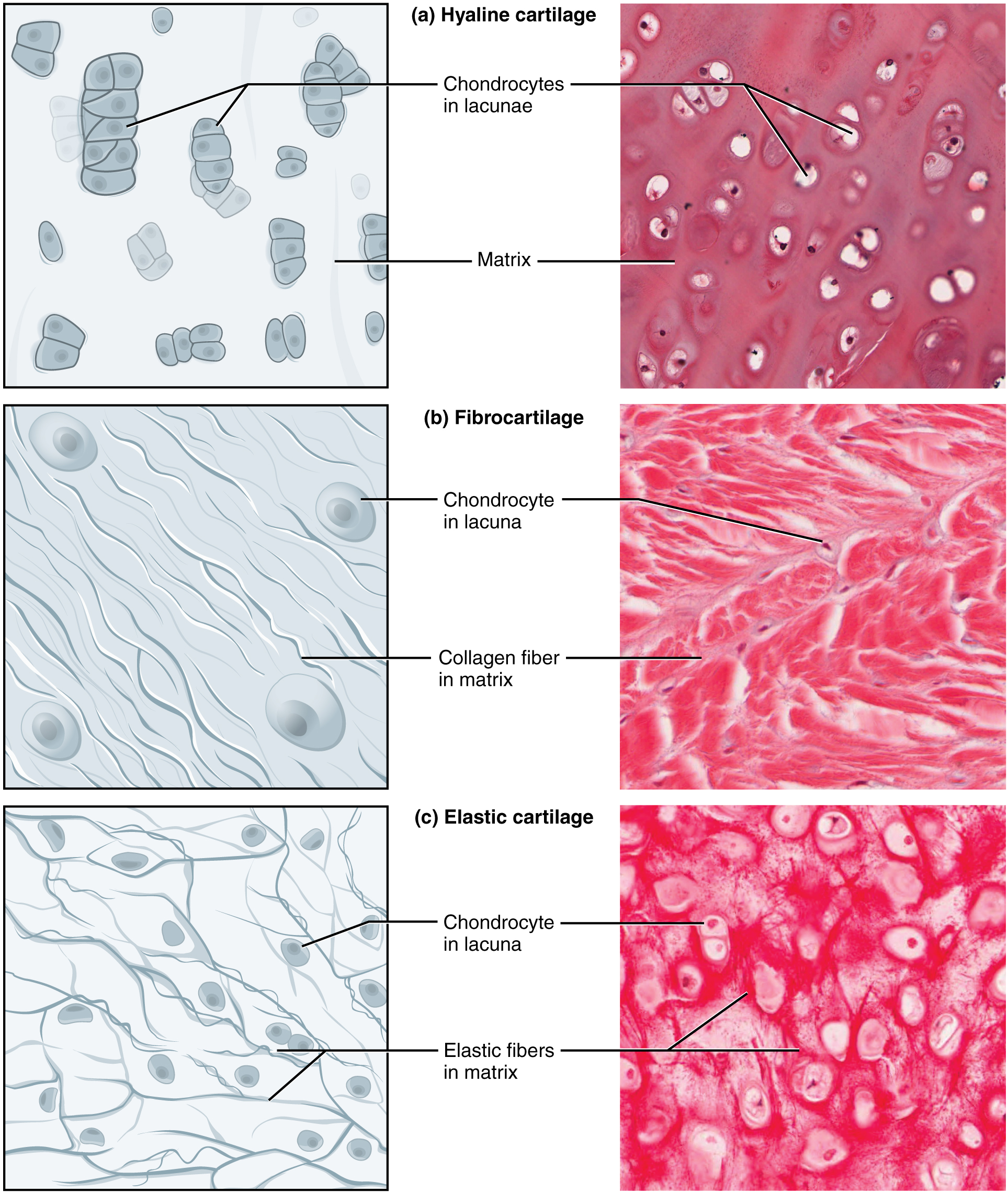
Connective Tissue Supports and Protects · Anatomy and Physiology
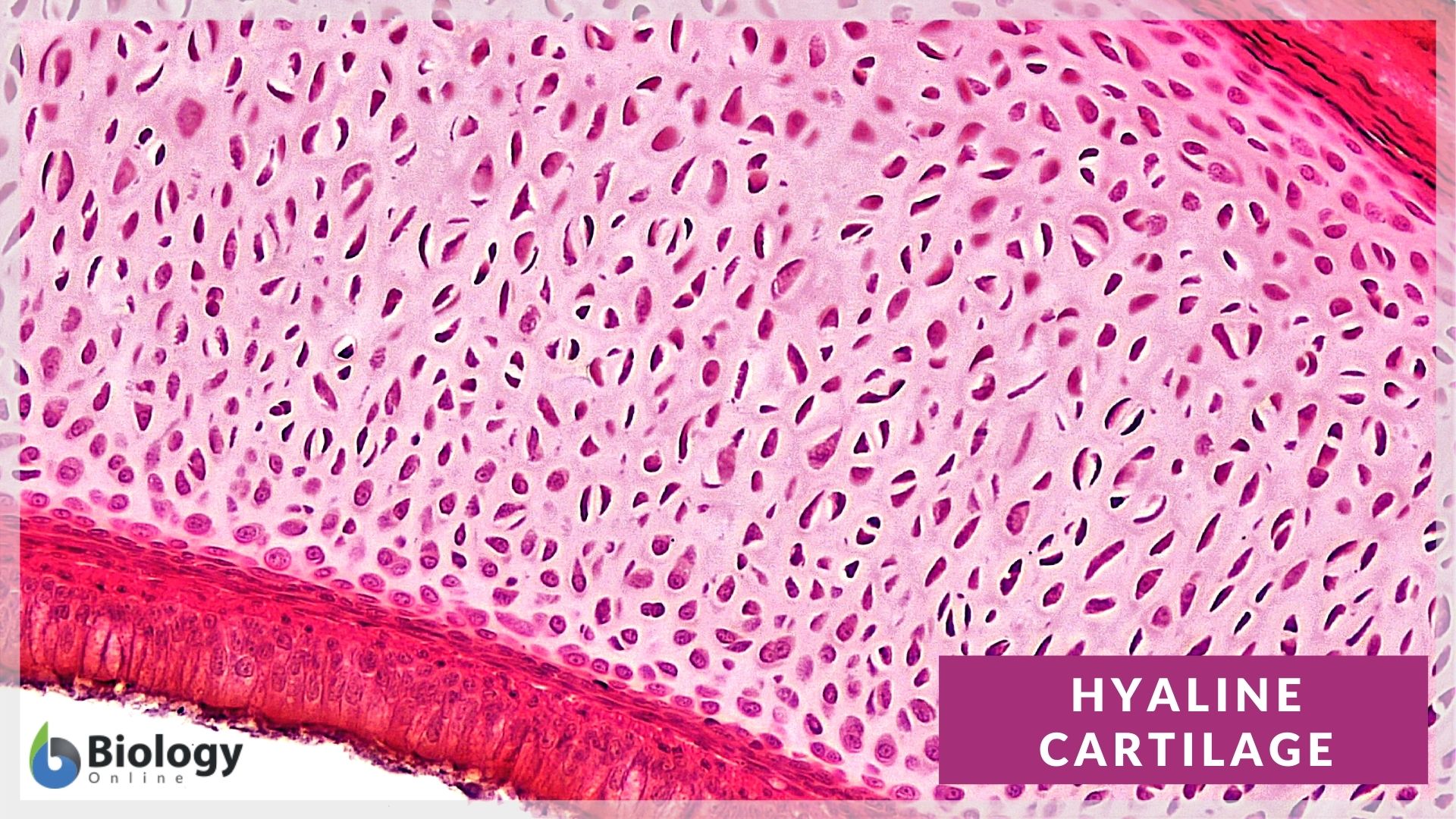
Hyaline cartilage Definition and Examples Biology Online Dictionary

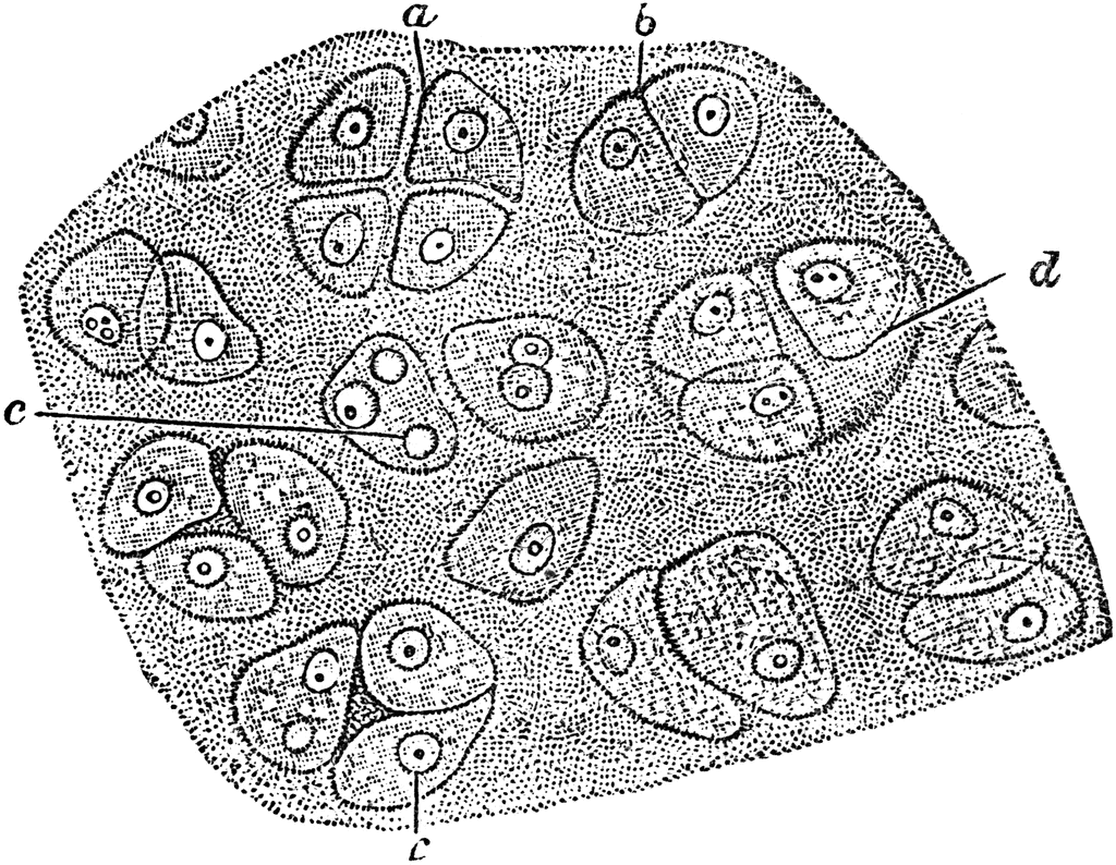
How to Draw Hyaline Cartilage Simple and easy steps Biology Exam
Hyaline Cartilage Cells ClipArt ETC
Hyaline Cartilage ClipArt ETC

Schematic drawing of articular (hyaline) cartilage containing abundant
Cartilage types a)Hyaline Cartilage Cartilage System

Illustrations Hyaline Cartilage General Histology

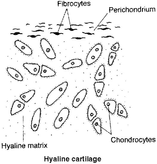
Hyaline Cartilage Labeled Diagram

Hyaline Cartilage Drawing YouTube
A Type Of Cartilage Found On Many Joint Surfaces;
Replaced By Bone Tissue As The Organ Grows In Length Epiphysis Wide Section At Each End.
Web During Embryonic Development, Hyaline Cartilage Serves As Temporary Cartilage Models That Are Essential Precursors To The Formation Of Most Of The Axial And.
Formed By The Process Of Chondrogenesis, The Resulting.
Related Post: