Drawing Of Esophagus
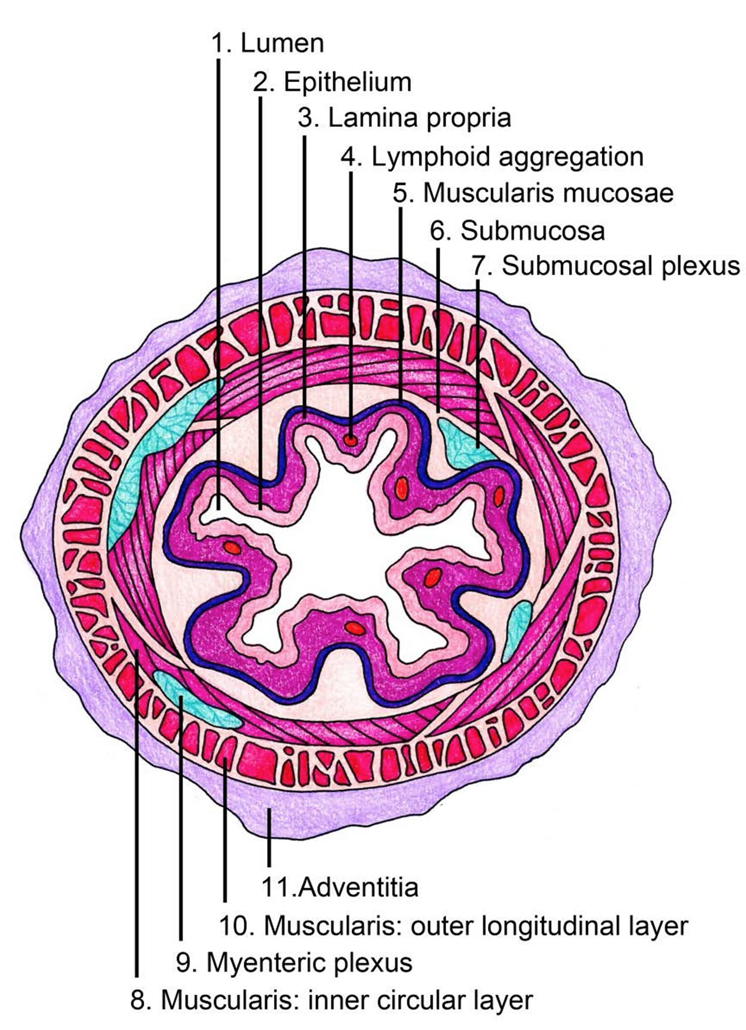
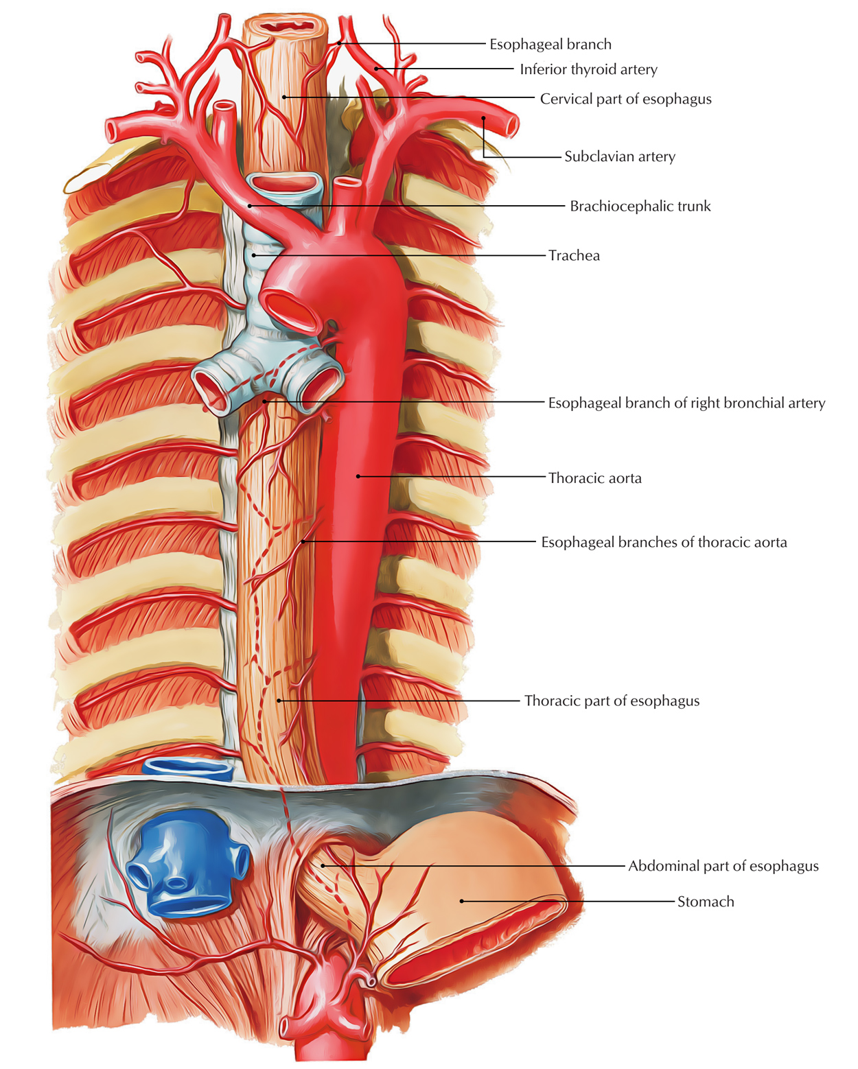
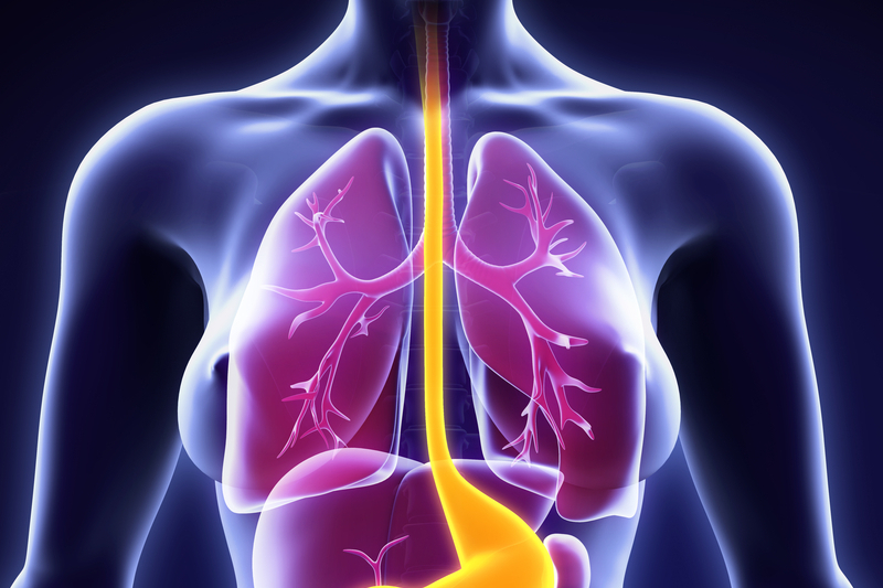
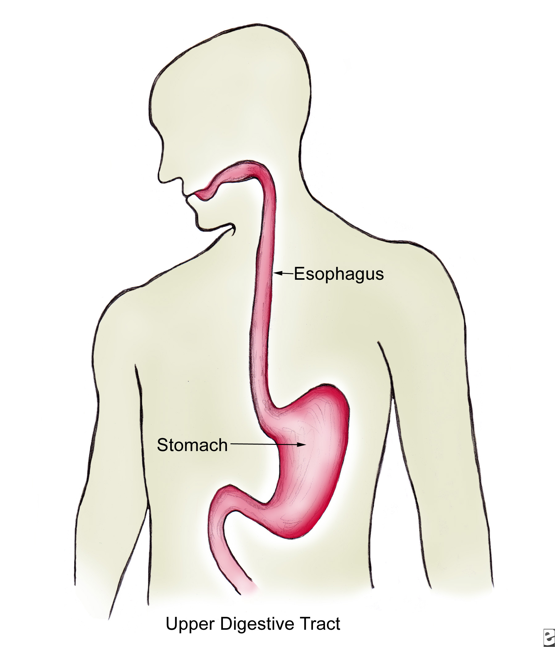
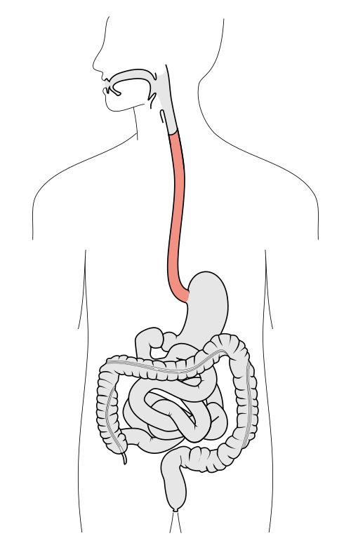
Drawing Of Esophagus - Web draw the esophagus. Web the esophagus is made up of four layers of tissue. Web the esophagus ( american english) or oesophagus ( british english, see spelling differences; Web do you want to get esophagus histology slide drawing tutorial? If left untreated, this condition can become very uncomfortable, causing. At 20x magnification, we can see each of the layers more clearly. Web the esophagus is a muscular tube about ten inches (25 cm.) long, extending from the hypopharynx to the stomach. Web in this section, you will examine the anatomy and functions of the three main organs of the upper alimentary canal—the mouth, pharynx, and esophagus—as well as three associated accessory organs—the tongue, salivary glands, and teeth. Newest results human digestive system woman anatomy diagram a medical anatomy diagram of a woman showing the human digestive system stomach in human body healthy stomach inside man body vector. Different layers of the esophagus. Web browse 236 drawing of esophagus stock photos and images available, or start a new search to explore more stock photos and images. Editable stroke healthy throat linear icon. Web the esophagus (oesophagus) is a 25 cm long fibromuscular tube extending from the pharynx (c6 level) to the stomach (t11 level). Web your esophagus is a hollow, muscular tube that. Web the esophagus is a muscular tube about ten inches (25 cm.) long, extending from the hypopharynx to the stomach. Esophagus drawing stock photos are available in a variety of sizes and formats to fit your needs. The throat includes the esophagus; It should be fairly narrow, about 1/5 the width of your model's neck. The esophagus lies posterior to. Esophagus drawing stock photos are available in a variety of sizes and formats to fit your needs. Web l isa thornton was heavily pregnant and in her early 30s when she noticed the feeling of a blockage in her oesophagus, the muscular food pipe that connects the mouth to the stomach. Oral cavity, pharynx, esophagus in good health. Colon, also. Web do you want to get esophagus histology slide drawing tutorial? Colon, also called the large intestine; You may follow the same but must try to draw better than this esophagus drawing. The mucosa of the esophagus consists of 3 main layers. Web anatomic drawings of the digestive system esophagus liver (right lobe) intrahepatic bile duct common bile duct gallbladder. Web esophagitis is an inflammation of the lining of the esophagus, the tube that carries food from the throat to the stomach. Web the esophagus ( american english) or oesophagus ( british english, see spelling differences; Here in this section i am going to share esophagus slide image drawing with you. Muscles in your esophagus propel food down to your. (o)esophagi or (o)esophaguses ), colloquially known also as the food pipe, food tube, or gullet, is an organ in vertebrates through which food passes, aided by peristaltic contractions, from the pharynx to the. The food moves from the mouth into the esophagus, which carries it down into the stomach. Web draw the esophagus. Web the esophagus ( american english) or. Web the esophagus ( american english) or oesophagus ( british english, see spelling differences; At the end of the mouth, draw a small tube that extends straight down into the center of your model’s torso. Web the esophagus is a muscular tube about ten inches (25 cm.) long, extending from the hypopharynx to the stomach. The mouth the cheeks, tongue,. The esophagus lies posterior to the trachea and the heart and passes through the mediastinum and the hiatus, an opening in the diaphragm, in its descent from the thoracic to the abdominal cavity. There are three layers of the mucosa: It consists of muscles that run both longitudinally and circularly, entering into the abdominal cavity via the right crus of. Here in this section i am going to share esophagus slide image drawing with you. Video trending searches climbing convention family reunion human body rock hall robotic arm popular categories Web the esophagus is made up of four layers of tissue. If left untreated, this condition can become very uncomfortable, causing. Web all 4 layers have variations to their structure. Colon, also called the large intestine; It consists of muscles that run both longitudinally and circularly, entering into the abdominal cavity via the right crus of the diaphragm at the level of the tenth thoracic vertebrae. You may follow the same but must try to draw better than this esophagus drawing. If left untreated, this condition can become very uncomfortable,. Windpipe, also known as the trachea; Healthy stomach inside man body vector illustration. Newest results human digestive system woman anatomy diagram a medical anatomy diagram of a woman showing the human digestive system stomach in human body healthy stomach inside man body vector. Esophagus drawing stock photos are available in a variety of sizes and formats to fit your needs. Web the esophagus (oesophagus) is a 25 cm long fibromuscular tube extending from the pharynx (c6 level) to the stomach (t11 level). The order of these layers from the inside out are: A medical anatomy diagram of a woman showing the human digestive system. If left untreated, this condition can become very uncomfortable, causing. It consists of muscles that run both longitudinally and circularly, entering into the abdominal cavity via the right crus of the diaphragm at the level of the tenth thoracic vertebrae. There are three layers of the mucosa: Web do you want to get esophagus histology slide drawing tutorial? Web draw the esophagus. At 20x magnification, we can see each of the layers more clearly. Here in this section i am going to share esophagus slide image drawing with you. Web browse 236 drawing of esophagus stock photos and images available, or start a new search to explore more stock photos and images. You may follow the same but must try to draw better than this esophagus drawing.
The Mouth, Pharynx, and Esophagus Biology of Aging
The Human Esophagus Functions and Anatomy and Problems
![]()
Esophagus Anatomy, sphincters, arteries, veins, nerves Kenhub

Anatomy Of The Esophagus

E.3. Esophagus
Esophagus Earth's Lab
Esophagus Facts, Functions & Diseases Live Science
The esophagus Structure of the esophagus

Esophagus / Endoscopy — High Plains Surgical Associates
Esophagus Libre Pathology
Web Esophagitis Is An Inflammation Of The Lining Of The Esophagus, The Tube That Carries Food From The Throat To The Stomach.
These Layers Are Similar All Throughout The Whole Digestive Tract.
Voice Box, Also Known As The Larynx;
Problems With The Esophagus Include Acid Reflux And Gerd.
Related Post: