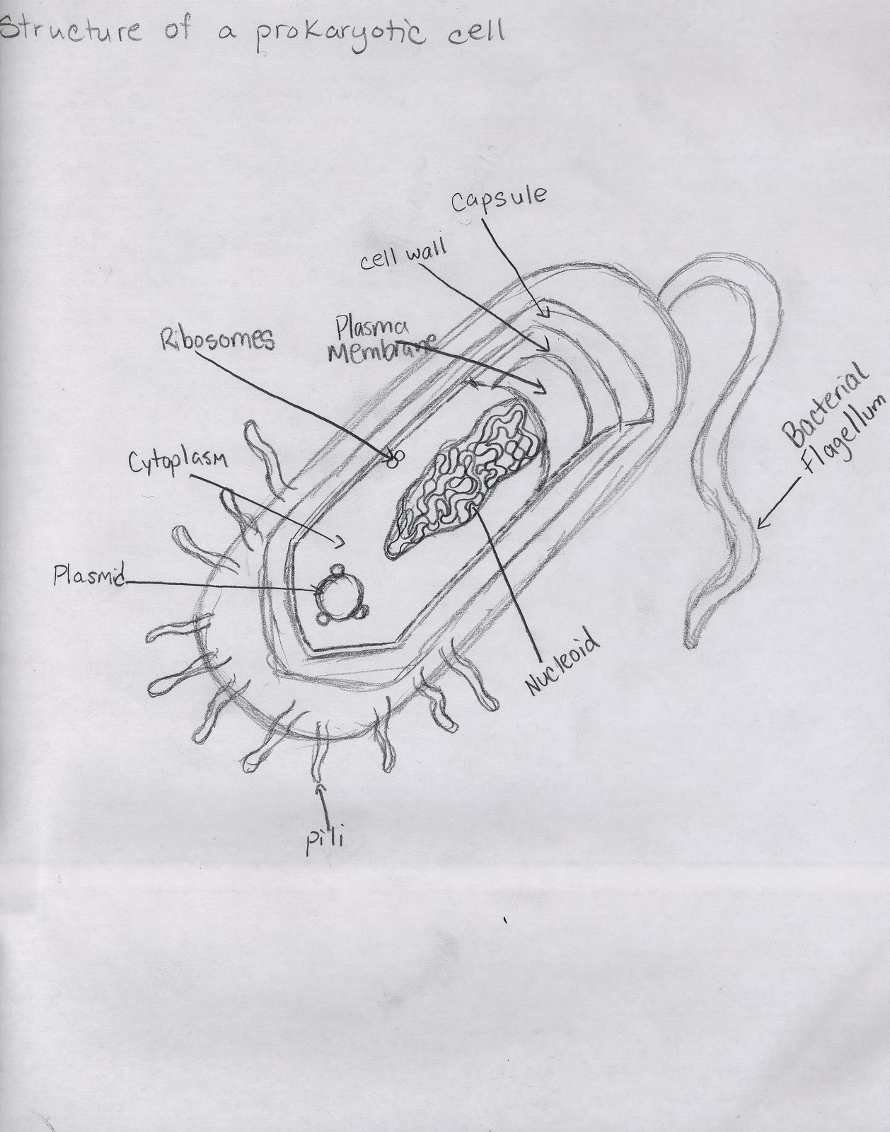
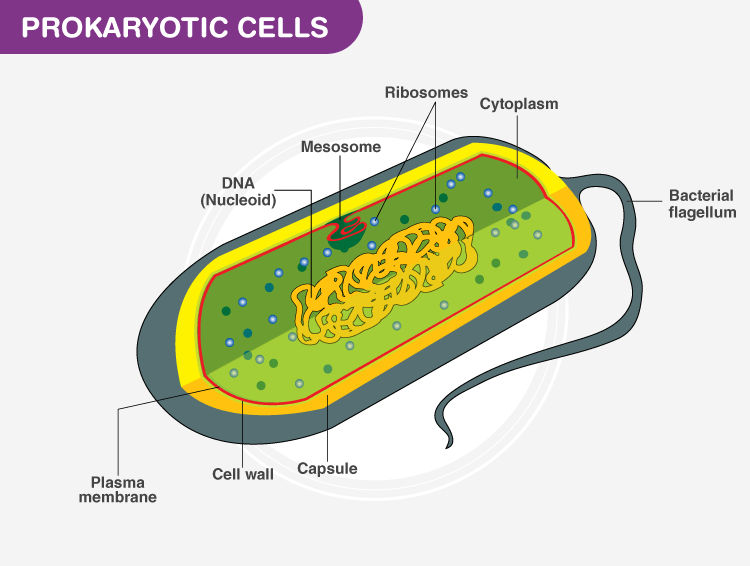
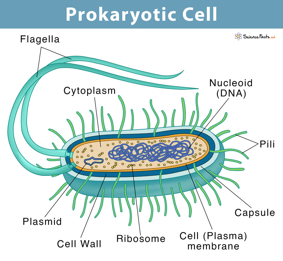
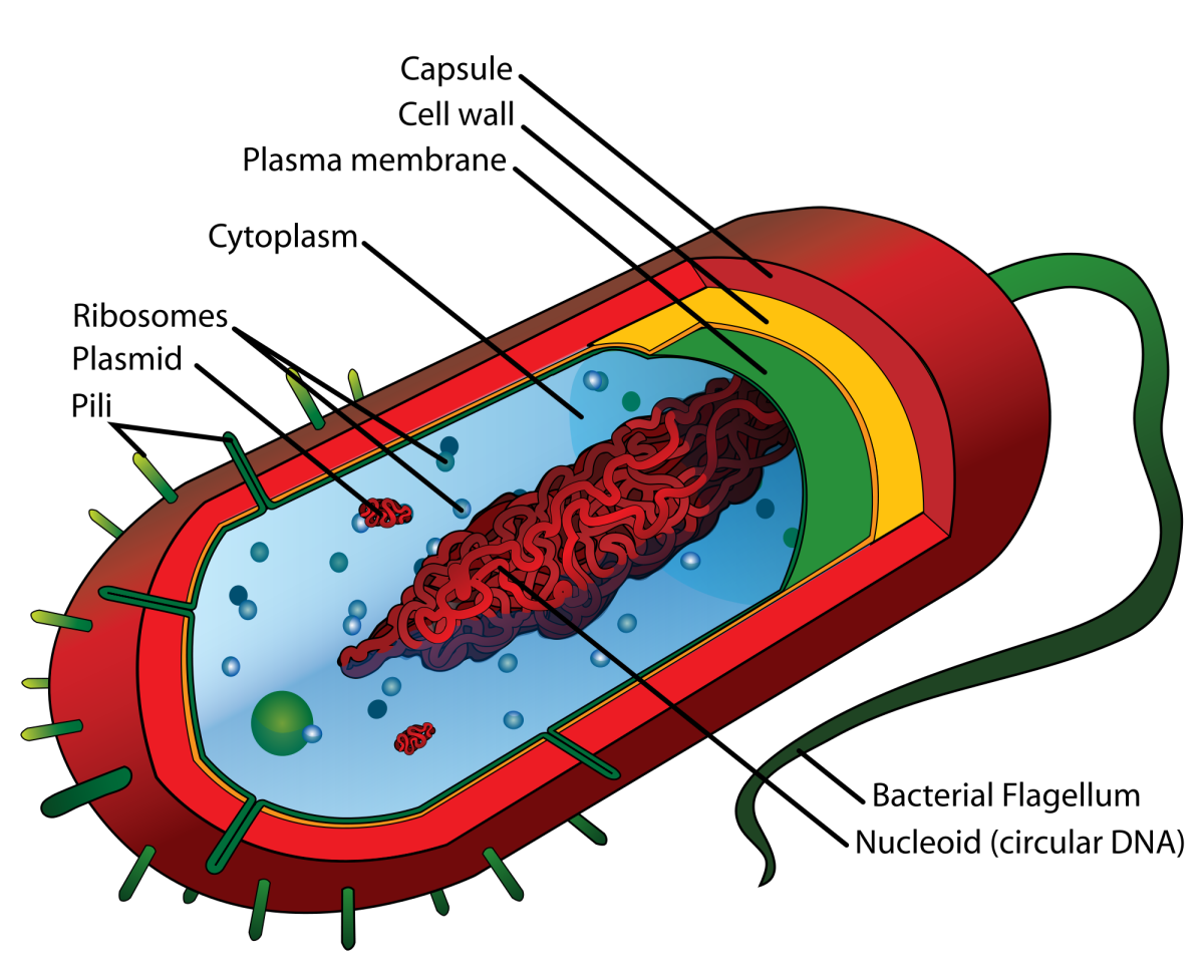
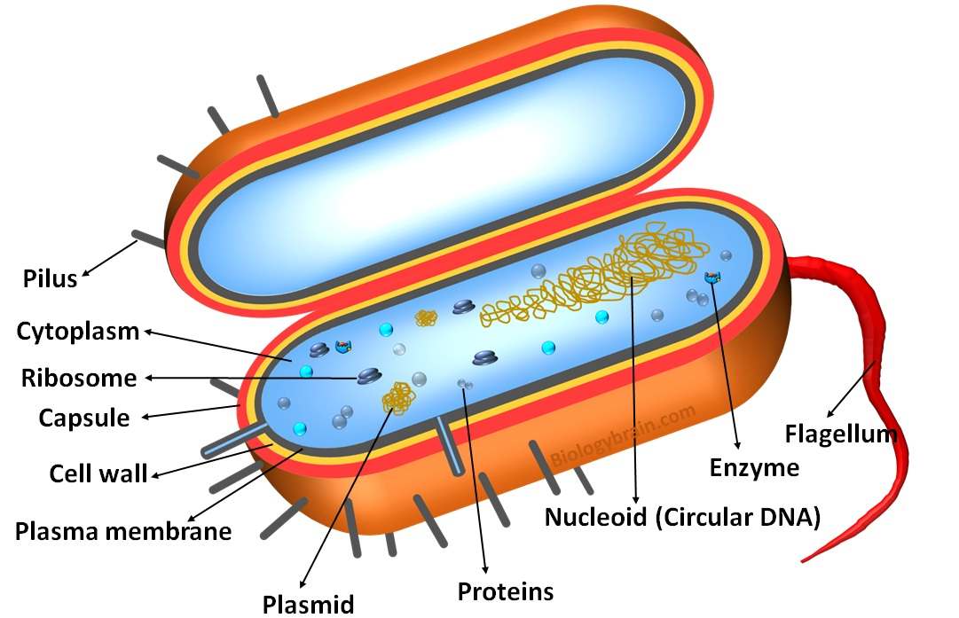
Drawing Of A Prokaryotic Cell
Drawing Of A Prokaryotic Cell - The structure called a mesosome was once thought to be an organelle. 2.2.2 annotate the diagram from 2.2.1 with the. The most common shapes are. Web most prokaryotic cells are much smaller than eukaryotic cells. Although they are tiny, prokaryotic cells can be distinguished by their shapes. Common prokaryotic cell is a bacterial cell. The prokaryotic cell diagram given below represents a bacterial cell. General structure of a prokaryotic cell: Web prokaryotic cells 2.2.1 draw and label a diagram of the ultrastructure of escherichia coli (e. Coli) as an example of a prokaryote. Prokaryotic dna is found in a central part of the cell: Coli) as an example of a prokaryote. Dna in eukaryotic cells is found inside. Web most prokaryotes have a cell wall outside the plasma membrane. Web prokaryotic and eukaryotic cells plasma membrane and cytoplasm google classroom structure and function of the plasma membrane and cytoplasm of cells. Many also have a capsule or slime layer made of polysaccharide. As i go, i give tips on drawing the various structures. Our body has over 100 trillion bacterial cells. Dna in eukaryotic cells is found inside. Web prokaryotic cells 2.2.1 draw and label a diagram of the ultrastructure of escherichia coli (e. Our body has over 100 trillion bacterial cells. Although they are tiny, prokaryotic cells can be distinguished by their shapes. This figure shows the generalized structure of a prokaryotic cell.all prokaryotes have. Most have peptidoglycan cell walls and many have polysaccharide. Dna in eukaryotic cells is found inside. These cells are very minute in size 0.1 to 5.0 μ m. Web most prokaryotes have a cell wall outside the plasma membrane. Web prokaryotic and eukaryotic cells plasma membrane and cytoplasm google classroom structure and function of the plasma membrane and cytoplasm of cells. This figure shows the generalized structure of a. Web diagram of a typical prokaryotic cell. Figure 22.10 the features of a typical prokaryotic cell are shown. These cells are very minute in size 0.1 to 5.0 μ m. It depicts the absence of a true nucleus and the presence of a flagellum. Prokaryotes often have appendages (protrusions) on their. Recall that prokaryotes are divided. As i go, i give tips on drawing the various structures. As organized in the three domain system, prokaryotes. Web i am demonstrating the colorful diagram of prokaryotic cells step by step which you can draw very easily. Although they are tiny, prokaryotic cells can be distinguished by their shapes. Web i draw a bacterial cell to show you how. Web prokaryotic and eukaryotic cells plasma membrane and cytoplasm google classroom structure and function of the plasma membrane and cytoplasm of cells. As organized in the three domain system, prokaryotes. 2.2.2 annotate the diagram from 2.2.1 with the. The structure called a mesosome was once thought to be an organelle. Prokaryotic dna is found in a central part of the. 2.2.2 annotate the diagram from 2.2.1 with the. Coli) as an example of a prokaryote. Common prokaryotic cell is a bacterial cell. Figure 22.10 the features of a typical prokaryotic cell are shown. The prokaryotic cell diagram given below represents a bacterial cell. These neat, well labelled and colorful diagrams will make your. Web prokaryotic and eukaryotic cells plasma membrane and cytoplasm google classroom structure and function of the plasma membrane and cytoplasm of cells. Prokaryotes often have appendages (protrusions) on their. Genetic material is not enclosed by a nuclear membrane. This figure shows the generalized structure of a prokaryotic cell.all prokaryotes have. Web i am demonstrating the colorful diagram of prokaryotic cells step by step which you can draw very easily. The prokaryotic cell diagram given below represents a bacterial cell. Web most prokaryotic cells are much smaller than eukaryotic cells. As organized in the three domain system, prokaryotes. Prokaryotic dna is found in a central part of the cell: These neat, well labelled and colorful diagrams will make your. Web prokaryotic and eukaryotic cells plasma membrane and cytoplasm google classroom structure and function of the plasma membrane and cytoplasm of cells. This figure shows the generalized structure of a. The structure called a mesosome was once thought to be an organelle. Dna in eukaryotic cells is found inside. Coli) as an example of a prokaryote. Many also have a capsule or slime layer made of polysaccharide. Although they are tiny, prokaryotic cells can be distinguished by their shapes. Figure 22.10 the features of a typical prokaryotic cell are shown. Web diagram of a typical prokaryotic cell. Web prokaryotic cells 2.2.1 draw and label a diagram of the ultrastructure of escherichia coli (e. Web most prokaryotic cells are much smaller than eukaryotic cells. The prokaryotic cell diagram given below represents a bacterial cell. As i go, i give tips on drawing the various structures. The most common shapes are. Web all prokaryotic cells are encased by a cell wall.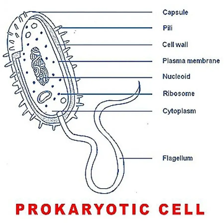
Simple Prokaryotic Cell Diagram
Cell Types and Structure Structure of Prokaryotic Cell

Prokaryotic Cells Labelled Diagram DIAGRAM

Prokaryotic Cell Diagram With Labels General Wiring Diagram

Prokaryotic Cell Structure, Characteristics & Function

3.3 Unique Characteristics of Prokaryotic Cells Biology LibreTexts
Prokaryotic Cells Definition, Structure, Characteristics, and Examples
Prokaryotic Cell Definition, Examples, & Structure
Prokaryotic Cell Structure A Visual Guide Owlcation
Labeled Prokaryotic Cell Diagram, Definition, Parts and Function
Web Most Prokaryotes Have A Cell Wall Outside The Plasma Membrane.
Most Have Peptidoglycan Cell Walls And Many Have Polysaccharide.
General Structure Of A Prokaryotic Cell:
2.2.2 Annotate The Diagram From 2.2.1 With The.
Related Post: